Is it time we start saying the E word?
Extinction is a dire word for an extreme event. There has been five mass extinctions in our planets history due to either asteroid impacts, climate change caused by volcanism and the shifting of tectonic plates. The sixth mass extinction is being fueled by you and me. Mass extinctions are relatively short periods of history during which a significant quantity of species die off beyond the standard background extinction (normal process of a species dying out and is usually a natural occurrence).
Ordovician Silurian Extinction
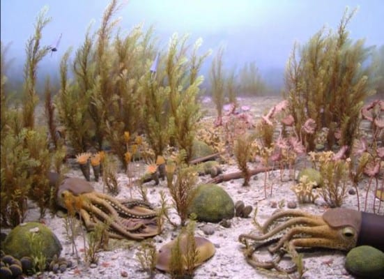
The Ordovician Silurian Extinction was actually two extinction in close proximity that took place 440-450 million years ago, killing off over 60% of all marine species. Possible causes:Weathering: heavy weathering of the Appalachian Mountains out-weighed volcanism, resulting in abnormally large CO2 sequestering, which lead to the development of a rapid ice age.
Gamma Ray Burst: this is a speculative hypothesis, but a gamma ray burst from a hypernova within 6,000 light years could have irradiated Earth. Within 10 seconds of the burst, half of the ozone layer would have been destroyed, bathing the planet in ultraviolet radiation.
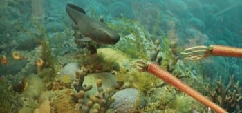
Late Devonian Extinction
A series of extinctions 360-375 million years ago, removed 70% of all species. Major environmental changes including frequent sea level changes, anoxic ocean bottoms, abnormally large carbon burials all contributed to the extinction; however it is not clear what caused the environmental changes in the first place. Possible causes:Plant evolution – Devonian sough rapid evolution of plants, in the early Devonian the tallest plants were 30 cm but by late Devonian plants escalated to 30 m (90 feet). Archaeopteris forests quickly covered the globe.
Effect on weathering – these plants contributed to heavy weathering as plant roots broke up soil/rock releasing huge quantities of nutrients in to the rivers and the ocean, the ecology of the time was not able to absorb the nutrients subsequently leading to anoxia.
Effect on CO2 – the greening of the planet absorbed CO2 leading to a permanent chilling to the planet.
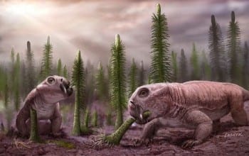 Permian-Triassic extinction
The Permian-Triassic extinction, which took place 251.4 million years ago, was the Earth’s largest, with 96% of all marine species and 70% of land species dying off. It’s also the only known mass extinction of insects. Possible causes:Methane Hydrate Gasification: the ocean sea-floor contains large amounts of methane hydrates, if those were to be released by a temperature change, the global temperature would increase by 6 degree Celsius (10.8F).
Flood Basalt Eruptions: Siberian Traps was one of the largest volcanic eruptions in Earth’s history, with over 2 million square kilometers covered in lava.
Impact Event: Major asteroid collision with Earth has been proposed as a possible cause but no large impact crater has been located within the right time-period; however, if the asteroid impacted an ocean, the crater would have been destroyed by “conveyor belt” sea-floor spreading. The oldest sea-floor is only 200 million years old.
Triassic-Jurassic Extinction
205 million years ago. 23%of all families and 48% of all genera went extinct. About half of the species o Earth were wiped out, including non-dinosaurian archosaurs (other than crocodiles), some therapids, and many large amphibians. Because of this, the reign of the dinosaurs officially began, most of their competition having been wiped out. This event happened fairly quickly on a geologic time scale (less than 10,000 years), right before the supercontinent Pangea began to break apart. There are several theories as to why this extinction happened, but nothing has been concretely found to determine the exact cause. The leading theories are gradual climate change, sea-level fluctuations, pulse of ocean acidity, asteroid impact, or massive volcanic eruptions (specifically from the Central Atlantic Magmatic Province, or CAMP). The reason could be any combination of these factors.
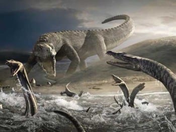
Cretaceous Tertiary event
The Cretaceous Tertiary event, taking place 70-65 million years ago, killed off 75% of all species, including the dinosaurs. This extinction cleared the way for mammals and birds, where before dinosaur were the dominant land species. Avian dinosaur fossils are the only ones found after the K-T boundary. Possible causes:Impact Event: an asteroid 10-15 km or 6-9 miles in diameter impacted Earth at Chicxulub, in Mexico’s Yucatan Peninsula. The explosion would have been a billion times larger than the nuclear bomb dropped on Hiroshima.
Flood Basalt Eruption: Deccan Traps flood basalt erupted for 800,000 years during the K-T boundary period, it would have exacerbated the devastation caused by the asteroid impact and would have slowed down the recovery of organisms.
An article from New Scientist reports on a study by Chris Thomas at the University of Leeds, UK, and colleagues, evaluating the impact on species of mild, moderate and severe levels of predicted climate change.Mr. Thomas is quoted as saying: “The absolutely best case scenario – which in my opinion is unrealistic – with the minimum expected climate change and all of the species moving completely into new areas which become suitable for them, means we end up with an estimate of nine per cent facing extinction,” This best case scenario means that 1 million species would go extinct assuming there are 10 million species in existence.
Many studies have looked at individual extinctions, while this study looks at migrations of species in a warming world. Thomas says the effects of climate change should be considered as great a threat to biodiversity as the “Big Three” – habitat destruction, invasions by alien species and overexploitation by humans.
Thomas states that his study overturns the belief that “climate change might simply result in the reassembling of species around the planet, without them dying out”.
Thomas and colleagues around the world statistically modelled the climates in which each of the 1103 species considered currently live. Whole groups of plant and animal species confined to a particular region, for example, the Amazon, were evaluated.Endangered species would have been included among these, as well as more common species, so Thomas says there is no reason to suppose that the organisms selected are unrepresentative of species generally.
A very important video below.
“We’ve been talking about rising sea levels and extreme weather events,” said author Thom Hartmann. ”Now we need to talk about extinctions.”Filmmaker Leila Conners and Producers George DiCaprio, Earl Katz, and Mathew Schmid have joined with Hartmann and writer/researcher Sam Sacks to produce a new short film – “Last Hours” – about the most extreme possible outcome of global warming.
Our planet has nearly died before. Five times in the deep geologic history of planet Earth, massive quantities of greenhouse gases have been released through the Earth’s crust. One was provoked by a meteorite strike, others by tectonic and volcanic activity. In each case, these massive releases of greenhouse gases warmed the planet enough to cause global mass extinctions – the death of more than half of all life.
“Last Hours” is a film that explores the possibility that we could be close – centuries or perhaps just decades -to tipping points that could lead to a sixth mass extinction, and relies on input from some of the world’s leading scientists.
The nail in our coffin? Methane.
Of the many tipping elements in the Arctic, that with potentially greatest consequences if perturbed is the vast methane deposit. Methane is a greenhouse gas. A molecule of methane has 20 times the greenhouse effect of a CO₂ molecule, and the release of methane has been linked to climatic transitions along the history of planet Earth.The Arctic contains vast reserves of methane stored as methane hydrate, a gel-like substance formed by methane molecules trapped in frozen water. The methane hydrate deposits are estimated at between 1,000 and 10,000 Gigatons (109 tons) of CO₂-equivalents as methane, much of which is present in the shallow sediments of the extensive Arctic shelves. This amount of greenhouse gas is several times the total CO₂ release since the industrial revolution.
Even moderate (a few degrees C) warming of the overlying waters may change the state of methane from hydrates to methane gas, which would be released to the atmosphere. If this release is gradual, methane will add a greenhouse effect to the atmosphere. This will only be temporary, as it will be oxidised to CO₂, with a decline in the greenhouse effect of 20-fold per unit carbon.
However, if the state shift is abrupt it may lead to a massive release of methane to the atmosphere, which could cause a climatic jump several-fold greater than the accumulated effect of anthropogenic activity.
Science Mag illustrates the venting of methane from the Siberian Eastern Shelf.
Remobilization to the atmosphere of only a small fraction of the methane held in East Siberian Arctic Shelf (ESAS) sediments could trigger abrupt climate warming, yet it is believed that sub-sea permafrost acts as a lid to keep this shallow methane reservoir in place. Here, we show that more than 5000 at-sea observations of dissolved methane demonstrates that greater than 80% of ESAS bottom waters and greater than 50% of surface waters are supersaturated with methane regarding to the atmosphere. The current atmospheric venting flux, which is composed of a diffusive component and a gradual ebullition component, is on par with previous estimates of methane venting from the entire World Ocean. Leakage of methane through shallow ESAS waters needs to be considered in interactions between the biogeosphere and a warming Arctic climate.
A must see video below, have your medication of choice handy as you will need it. You can download google 3d imaging from You Tube or from Methane Tracker, but my pc would not allow me to do it. Skip to 3:09 as the scientists took a while getting the graphics up and running.
This is absolutely bone chilling.
Video 1 of 2. This is a short intro to the Unified Methane Layers functionality on methanetracker.org.
Basically you’re looking at two layers per day (0-12z and 12-24z) that contains only the methane over 1950ppb of all 100 layers from IASI for each (am and pm). Those two layers are shown at 6am (0-12z) and 6pm (12-24z) each day. The visualization using the Google Earth plugin allows you to pick a from and to data range that makes it easier to visualize the actual geographical scope of the venting episode, as well as identify specific geographic locations and dates to run a more detailed analysis using the “Individual Methane Layers” functionality.


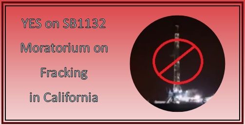 Key votes will be held this week on California Senate Bill 1132, which imposes a moratorium on hydraulic fracturing, or fracking as it commonly known. The fracking process poses many unacceptable public policy risks. These include contaminating water supplies; degrading public health; disproportionately affecting low income families and communities of color; using scarce water supplies in drought-stricken states; causing earthquakes; and harming wildlife and habitat fragmentation. If the bill fails, the legislative process toward moratorium must restart next January.Please join us for a blogathon May 19-23 in a campaign to tell lawmakers to support this bill. This is a coordinated effort with a coalition of more than a dozen NGOs, including Earth Works, Sierra Club, and Center for Race, Poverty and the Environment.